KOR Request a Quote
Scanning Electron Microscope · COXEM

Scanning Electron Microscope (SEM)

From tabletop SEMs to high-performance SEMs with up to 500,000× and large-area nanoparticle analysis, plus sample preparation equipment. The new NanoStation 5.0 software shows SE · BSE images live on a quad-split screen.

500,000×Max magnification (CX series)
< 3 nmSpatial Resolution
3 sAutofocus
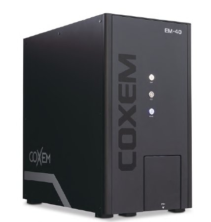
COXEM EM-40, CX-200K, CX-300

Product Lineup

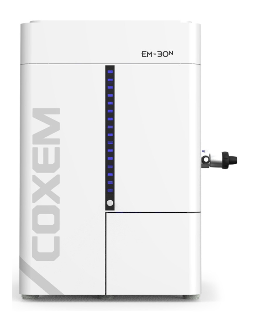
EM-30N
Tabletop SEM · EM Series

EM-30N

A tabletop SEM ready to use on your desk. With a tungsten filament electron gun and turbo pump vacuum system, it observes up to 150,000×.

Max magnification 150,000× · spatial resolution < 5 nm
Auto focus · auto brightness/contrast · auto start
X · Y 35 mm stage, tilt 0–45°
Dual-split screen · ChamberCam · Panorama v1
BSE · EDS · EBSD · STEM options
EM-40
Tabletop SEM · The Next Level of EM Series

EM-40

A tabletop SEM for your desk. It observes up to 250,000×, higher than the previous EM-30N, and is the world’s first tabletop SEM with EBSD.

New NanoStation 5.0 GUI
High-speed scanning up to 13 fps (ULTRA / TV / SLOW)
X · Y · Z 3-axis motorized stage
Variable pressure (10 / 20 / 30 Pa)
Compact EBSD · EDS high-speed particle analysis
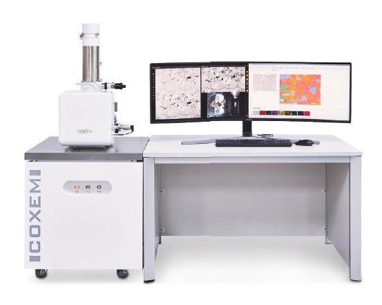
CX-200K
The Standard, Yet Powerful

CX-200K

A standard high-performance SEM with up to 500,000× magnification. A self-diagnosis system and 5-axis motorized stage make analysis stable and convenient.

New NanoStation 5.0 GUI
Max magnification 500,000×
Self-diagnosis system · optimized imaging
5-axis motorized stage
Air-isolation module, EDS · EBSD · Coolstage · STEM options
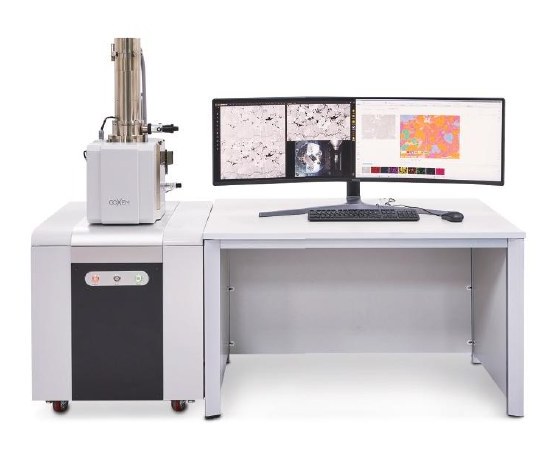
CX-300
The Advanced Innovation

CX-300

An advanced SEM for large-area analysis, providing a solution that systematically classifies the size, distribution and composition of nanoparticles on membrane filters.

Large-area design (stage X · Y 100 mm)
Convenient operation with NaviCam · ChamberCam
1-second auto brightness · contrast, quad-split screen
Fine dust · engine oil · microplastics · mineral analysis

Key Functions · Options

NanoStation 5.0View SE · BSE · SE+BSE · NaviCam images live on a quad-split screen.
3-second autofocusOne-click autofocus greatly reduces analysis time.
Position memory · sequential magnification captureStores up to 8 analysis positions and automatically captures and saves images at specified magnifications.
Panorama v2Enhanced stitching precisely joins images of large samples.
Variable pressure (VP)Analyze biological samples and insulating materials without coating in 10 / 20 / 30 Pa modes.
EBSD · EDSSupports crystallographic analysis and high-speed particle analysis of mixed samples.
STEM · CoolstageAnalyze asbestos and tissue with the STEM detector, and observe hydrated samples without damage on the cooling stage.
Air-isolation ModuleAir-sensitive samples prepared in a glove box are transferred to the SEM and observed without exposure.
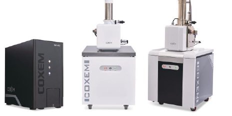
Model Comparison

Model EM-30NEM-40CX-200KCX-300
TypeTabletopTabletopStandardLarge-area Advanced
Electron GunTungsten filament (W)Tungsten filament (W)Tungsten filament (W)Tungsten filament (W)
Spatial Resolution< 5 nm< 5 nm< 3 nm< 3 nm
Photo magnification15× – 150,000×8× – 150,000×6× – 300,000×6× – 300,000×
Display magnification15× – 150,000×13× – 250,000×9× – 500,000×9× – 500,000×
Accelerating Voltage1 – 30 kV1 – 30 kV1 – 30 kV1 – 30 kV
Sample StageX 35 · Y 35 mm, Tilt 0–45°X 40 · Y 40 · Z 40 mm, 3-axis motorizedX 60 · Y 60 · Z 60 mm, R 360°, T -20–90°X 100 · Y 100 · Z 60 mm, R 360°, T -20–90°
DetectorSE, BSE*SE, BSE*SE, BSESE, BSE
Vacuum ModeHV, LV*HV, LV*, VP*HV, LV*, VP*HV, LV*, VP*
Vacuum SystemTurbo 80 L + Rotary 100 LTurbo 80 L + Rotary 100 LTurbo 80 L + Rotary 100 LTurbo 80 L + Rotary 100 L
Screen Layout2-split3-split3-splitQuad-split
Dimensions · Weight400 × 630 × 600 mm, 85 kg315 × 560 × 580 mm, 81.5 kg640 × 682 × 1,432 mm, 200 kg640 × 690 × 1,460 mm, 200 kg

* Options and common specs · Output formats JPEG, TIFF, BMP · Common options EBSD, STEM, Coolstage, NanofiberScanner

Observation Examples

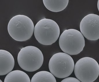
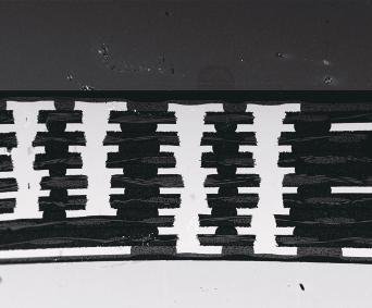
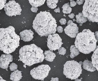
Metal powder, semiconductors, PCB, metal plate, nickel powder, mask sheet, solder balls, minerals, biological samples, etc.

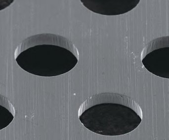
Metal Plate SEM image
Metal Plate
Solder Balls (Tin ball) SEM image
Solder Balls (Tin ball)
PCB SEM image
PCB
Nickel Powder SEM image
Nickel Powder

Sample Preparation Equipment

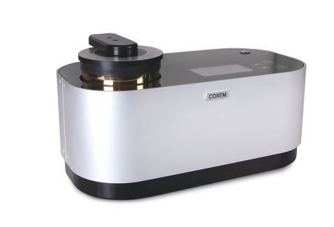
SPT-20
Ion Sputter Coater

SPT-20

Deposits a thin conductive film on samples to protect the surface and obtain sharper SEM images. One-touch LCD operation and current feedback for stable coating.

TargetAu, Pt, Pt-Pd (Ø50 mm)
ChamberØ100 mm
Max Ion Voltage3 kV
Ion Current0 – 9 mA
Coating Time10 – 300 s
Dimensions · Weight420 × 220 × 230 mm, 10 kg
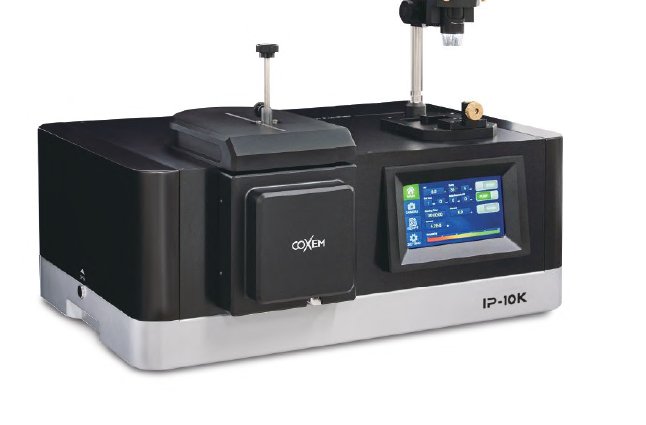
IP-10K
Ion Beam Polisher

IP-10K

Precisely polishes sample cross sections with an ion beam. It produces cleaner cross sections than mechanical polishing and handles sensitive samples with a cooling stage and air-isolation module.

Milling Rate1,000 µm/h (Si, 8 kV)
Ion Accelerating Voltage2 – 8 kV
Cooling Module-20 °C (Peltier)
Flat millingTilt 10° – 90°, Ø32 × 18 mm
Operation7-inch touch panel
WeightMain unit 45 kg

Tell us your samples and required magnification, and we will propose the right model and options

T. 042-361-9999 · E. lin-tech@lin-tech.co.kr
Request a Quote · Demo